Frontiers of Neurodegeneration Treatment: Targeting the Brain's Border Immune Checkpoints
Evidence level: Multiple Level 1-2 natural experiments (N=850,000); mechanistic support from randomized controlled trials and AI-validated biomarker studies
A 70-year-old Canadian whose birthdate fell on the eligible side of Ontario's January 1, 1946 vaccine-program cutoff carried a 2.0 percentage point lower absolute risk of dementia than those born just after the cutoff. The difference? Eligibility for the Zostavax shingles vaccine under Ontario's birthdate cutoff policy. That two-percentage-point gap, tracked across 464,000 Canadians over 5.5 years and published in *Lancet Neurology*, marks the fourth independent replication of the most clinically robust dementia-prevention signal in human data: peripheral immune interventions reaching deep into brain outcomes through mechanisms that bypass the blood-brain barrier entirely.
The pattern holds across four natural experiments spanning approximately 950,000 people in Wales, Australia, the United States, and now Canada, with remarkably consistent effect sizes: 17–20% reductions in dementia incidence, significantly stronger in women. The recombinant Shingrix vaccine showed a 17% increase in dementia-free time compared to Zostavax in a US study of 103,615 people—matching the clinical magnitude of monoclonal antibodies that target amyloid plaques directly but require intravenous infusion and carry brain-swelling risks. Yet no neurologist writes "shingles vaccine for brain protection" because until recently, we lacked the anatomy to explain how a shoulder injection against varicella-zoster virus could alter Alzheimer's disease progression.
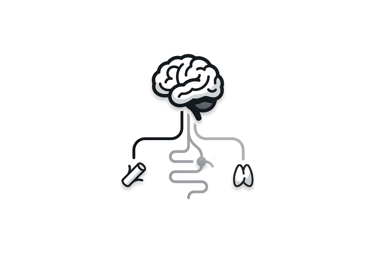
Three converging discoveries now provide that anatomy: meningeal lymphatic vessels reportedly draining brain waste to cervical nodes, the vagus nerve's inflammatory reflex shown modulatable in FDA-approved device trials, and thymus health scores quantified by AI across approximately 25,000 CT scans from 27,612 participants. Together, they reveal the brain operates not as an immune-isolated fortress but as the downstream terminus of an integrated periphery-to-parenchyma clearance system—one whose accessible checkpoints lie outside the skull.
Last April, Ground Truths covered the shingles-dementia epidemiology as a striking but mechanistically opaque correlation. This week's evidence clarifies the biology: three independent highways connect peripheral immune state to neurodegeneration outcomes, each now mapped at sufficient resolution to guide intervention design. The mechanistic convergence matters because it inverts therapeutic strategy—we've spent decades trying to drug the brain directly when the immune system's border control stations offer faster paths to human proof-of-concept.
The Lymphatic Highway
For a century, anatomists taught that the brain lacks lymphatic vessels, making waste clearance a mystery. The reportedly 2015 discovery of genuine lymphatic capillaries lining the dural sinuses—the venous channels between skull and brain—demolished that axiom. These vessels drain cerebrospinal fluid and brain interstitial waste to deep cervical lymph nodes, the same nodes that survey throat infections.
Inject fluorescent tracer into mouse brain parenchyma and it appears in cervical nodes within hours, carried by bulk flow through newly characterized routes: along arterial sheaths into brain tissue (the glymphatic influx, driven by arterial pulsation and active mainly during sleep), through the interstitial space where astrocyte water channels facilitate convective flow, then out along venous pathways to the subarachnoid space, across a thin mesothelial membrane that partitions cerebrospinal fluid compartments, and finally into dural lymphatics. The system clears amyloid-beta, tau, alpha-synuclein—the protein aggregates defining Alzheimer's and Parkinson's diseases—at rates that decline with age and correlate with cognitive trajectory.
Human translation is underway. Lymphatic-venous anastomosis surgery—creating artificial connections between deep cervical lymphatics and jugular veins—now operates at 30 centers in China for Alzheimer's and Parkinson's patients, with randomized trials launching in the United States. The logic: if drainage capacity limits waste clearance, bypass the bottleneck surgically. Cervical lymph nodes are palpable, biopsyable, imageable; interventions targeting them avoid blood-brain barrier penetration challenges that have sunk dozens of small-molecule drug candidates.
The Vagal Highway
The vagus nerve, a thumb-thick cable connecting brainstem to viscera, carries 80% afferent signals—the gut, heart, and immune organs report constantly to brain. Kevin Tracey's three-decade research program proved the reverse pathway matters equally: efferent vagal signals suppress peripheral inflammation through the "inflammatory reflex," a term he coined after observing that vagus nerve stimulation drops tumor necrosis factor levels within minutes. A sham-controlled randomized trial in *Nature Medicine* demonstrated that one minute daily of barely perceptible electrical stimulation—a device implanted alongside the neck vagus in a one-hour surgery—achieved FDA approval for refractory rheumatoid arthritis unresponsive to immunosuppressive drugs.
The mechanism: vagal efferents trigger acetylcholine release that binds alpha-7 nicotinic receptors on macrophages, silencing inflammatory cytokine transcription.
The brain connection runs through multiple channels. Vagal afferents synapse in brainstem nuclei that project to hypothalamus and limbic structures governing stress response and microglial activation state. Chronic low-grade inflammation—the "inflammaging" detectable by elevated C-reactive protein and interleukin-6—predicts dementia incidence in prospective cohorts. If vagal stimulation can dampen peripheral inflammation, and peripheral inflammatory state influences brain outcomes (as the shingles vaccine data prove), then vagus nerve modulation becomes a testable dementia-prevention strategy. Trials are starting for lupus and Crohn's disease; the neurodegeneration indication follows the same logic with stronger epidemiological backing.
The Thymic Highway
The thymus gland, tucked behind the sternum, shrinks with age—the involution process that turns spongy T-cell nurseries into fat. Two reportedly landmark *Nature* papers used AI to score thymus health from chest CT scans (a 0–100 scale derived from 4,096 image features, processing each 30-megabyte scan in under two seconds), then linked scores to outcomes across 27,612 people followed for more than 12 years. High thymus scores were associated with reduced mortality, with cardiovascular mortality showing a weighted hazard ratio of 0.53 across cohorts. Inflammation markers ran inverse: people with low thymus scores showed elevated C-reactive protein and interleukin-6, the same inflammatory signature that precedes dementia.
The molecular bridge from thymic involution to neurodegeneration emerged in reportedly recent studies of the neuroproteasome—a membrane-bound variant of the protein-shredding proteasome complex unique to neurons. Unlike cytosolic proteasomes that digest proteins internally, neuroproteasomes embed in the cell membrane, ingesting misfolded proteins from one side and ejecting peptide fragments externally. Block these neuroproteasomes (using inhibitors too large to cross membranes, leaving internal proteasomes intact) and tau aggregates form within hours—specifically the phosphorylated, insoluble filaments identical to Alzheimer's tangles. The genotype effect is dramatic: neurons carrying two copies of ApoE4, Alzheimer's strongest genetic risk factor, form tau aggregates after losing just 20% of neuroproteasome activity.

ApoE3 neurons require 60% loss; ApoE2 protective-variant neurons tolerate 85% loss before aggregating. ApoE physically binds neuroproteasomes at the membrane; the E4 variant reduces surface neuroproteasome levels by half compared to E3 in human postmortem hippocampus.
Thymic involution means fewer naïve T cells, skewed T-cell repertoires, chronic low-grade inflammation—an aged immune system that may inadequately clear peripheral infections (like varicella-zoster reactivation) and generates inflammatory signals that cross into brain via multiple routes. The shingles vaccine data show this matters clinically: the effect size is significantly larger in women (who have higher Alzheimer's incidence and stronger inflammatory responses) and in ApoE4 carriers, precisely the populations whose neurons run closest to the neuroproteasome failure threshold. Vaccination doesn't just prevent shingles; it recalibrates immune tone systemically. Studies in 3,884 participants from the U.S.
Health and Retirement Study found shingles vaccination associated with slower epigenetic aging (measured by DNA methylation clocks: PhenoAge, GrimAge, DundedinPACE) and reduced inflammation markers, effects durable beyond four years and strongest in the first three years post-vaccination when dementia-risk reduction also peaks.
The Therapeutic Inversion
The therapeutic inversion becomes clear: instead of designing molecules to cross the blood-brain barrier, clear amyloid plaques, and hope for cognitive benefit (the strategy that consumed 30 years and billions in failed trials), target the accessible immune checkpoints that gate whether inflammatory signals and waste products reach critical thresholds in brain. The checkpoints are mappable, measurable, and already clinically accessed. Vagus nerve stimulators exist as FDA-approved devices with established safety profiles; the indication expansion from epilepsy and depression to rheumatoid arthritis proves immune modulation works in humans, and the dementia trials require only adding cognitive outcomes to protocols already designed. Lymphatic-venous anastomosis surgery happens in 30 centers; randomized sham-surgery trials launching in the U.S.
will establish efficacy with far faster readouts than decades-long prevention trials. Thymus health scores derive from routine chest CT scans ordered for lung cancer screening in millions of people annually; the AI model runs in two seconds per scan and predicts mortality with effect sizes (hazard ratios 0.38–0.57) rarely seen in biomarker literature.
We have spent three decades banging on the front door—trying to force drugs through the blood-brain barrier to target proteins already aggregated in dying neurons.
The clinical evidence level matters. The shingles vaccine data represent Level 1–2 evidence: four independent natural experiments, birthdate-based eligibility creating quasi-randomization, approximately 950,000 total participants, 5–12 year follow-ups, consistent 17–20% effect sizes, dose-response relationships (Shingrix recombinant vaccine outperforms live Zostavax), and biological gradients (stronger effects in women and ApoE4 carriers where mechanisms predict greater vulnerability). No Alzheimer's drug candidate has remotely comparable human evidence for disease modification. The shingles vaccine takes two shots, and the epidemiological signal appears in mixed populations including people with established dementia.
The mechanistic convergence explains what seemed paradoxical: a peripheral viral vaccine reducing Alzheimer's dementia through pathways having nothing to do with varicella-zoster virus in brain. The vaccine's effect operates through systemic immune recalibration—dampening chronic inflammation, potentially improving thymic output or T-cell repertoire quality, reducing the inflammatory drive that pushes vulnerable neurons past neuroproteasome capacity. The signal propagates via lymphatic drainage (carrying inflammatory mediators from injection site to cervical nodes adjacent to brain-draining lymphatics), vagal afferents (sensing peripheral immune state and adjusting central inflammatory tone), and thymic function (if vaccination triggers transient immune activation that resets aging-related dysfunction, a mechanism proposed but not yet proven).
The Research Program
The research program writes itself. First: measure thymus scores, inflammatory biomarkers, and lymphatic drainage capacity (now quantifiable using gadolinium-enhanced MRI of cervical nodes) before and after shingles vaccination in ApoE4 carriers, tracking cognitive outcomes and neurodegeneration biomarkers (plasma p-tau217, neurofilament light) over five years. Second: test vagus nerve stimulation in mild cognitive impairment, using reportedly one-minute daily stimulation protocols in rheumatoid arthritis patients, with tau-PET and cognitive testing as endpoints. Third: randomize lymphatic-venous anastomosis surgery versus sham in early Parkinson's disease, where alpha-synuclein clearance via lymphatics offers a testable mechanism and motor outcomes provide faster readouts than dementia trials.
Fourth: determine whether handheld transcutaneous vagal stimulators (avoiding implant surgery) deliver sufficient inflammatory modulation to move dementia biomarkers, enabling population-scale prevention trials.
The border-checkpoint framework reframes neurodegeneration as fundamentally a failure of brain-immune integration—the progressive breakdown of waste clearance and immune surveillance systems that normally operate across scales from molecular (neuroproteasomes) through cellular (microglial activation) to organ-system (lymphatic drainage, vagal reflexes). Aging degrades all three highways simultaneously: thymic involution skews immune state, arterial stiffening reduces glymphatic flow, lymphatic vessel regression impairs drainage. The system has redundancy when young; by age 70, losing any one component tips others past compensation thresholds. ApoE4 carriers start with 50% lower neuroproteasome reserve; women show stronger inflammatory responses to immune challenges; these populations hit failure thresholds earlier, explaining the genetic and sex-specific epidemiology.
The implications extend beyond Alzheimer's. Parkinson's disease shows identical lymphatic-clearance deficits and responds to systemic inflammation reduction. Multiple sclerosis—once the paradigm autoimmune CNS disease—now appears driven partly by meningeal lymphatic dysfunction allowing autoreactive T cells access to brain. Even psychiatric conditions show immune-border signatures: depression correlates with low vagal tone (measurable as heart rate variability) and responds to vagus nerve stimulation in treatment-resistant cases. The brain-immune interface, far from being a specialized quirk of CNS neurobiology, may represent the control surface for a remarkable range of conditions.
We have spent three decades banging on the front door—trying to force drugs through the blood-brain barrier to target proteins already aggregated in dying neurons. The shingles vaccine data, replicated across four countries and approximately 950,000 people with effect sizes matching our best drugs, prove the back door works: peripheral immune interventions alter brain outcomes through highways mapped in sufficient detail to guide rational therapy design. The lymphatic vessels are there, draining to surgically accessible nodes. The vagus nerve is there, carrying inflammatory signals modulatable by FDA-approved devices. The thymus is there, its health scoreable from existing medical images and its function potentially restorable by interventions now moving toward human trials. The question is no longer whether peripheral immunity reaches brain—the anatomy and epidemiology prove it does.
The question is how fast we can translate these accessible, measurable, already-clinically-targeted checkpoints into the next generation of neurodegeneration trials, before another decade disappears into blood-brain barrier penetration attempts for targets that may not matter as much as the waste-clearance highways we only just discovered existed.